can human stomach acid dissolve bone
 What happens if you swallow a small poultry bone? Will it be digested or will it cause damage to the digestive system? - Quora
Warning: The NCBI website requires JavaScript to operate. Ingested bone fragment in the intestine: Two cases and a literature review Correspondence to: Selim Sözen, MD, Department of General Surgery, Medical Faculty, Namik Kemal University, 59000 Tekirdağ, Turkey. Phone: +90-282-2505500 Fax: +90-282-2509928AbstractIn general, ingested foreign bodies are excreted from the digestive tract without complications or morbidity. In adults, ingestion of foreign bodies often occurs in alcoholics and elderly with dentures. The most ingested foreign bodies are foods or their parts, such as fish bones or bone fragments and phytobezoars. Strange sharp bodies such as fish and chicken bones can lead to intestinal perforation and peritonitis. In the present report two cases, one of intestinal perforation and another of anal impact, both caused by ingested bone fragments. Complications due to ingested bone fragments are not common and preoperative diagnosis remains a challenge and should therefore be considered in susceptible cases. Basic tip: Ingested bone fragment can cause intestinal perforation anywhere in the syjunum on anal margin, obstruction and formation of fistula. An experienced doctor should suspect such conditions in the presence of certain predisposed factors, such as fast food and the use of dentures in the elderly, and should consider various surgical options. In the present report two cases, one of intestinal perforation and another of anal impact, both caused by ingested bone fragments. Complications due to ingested bone fragments are not common and preoperative diagnosis remains a challenge and should therefore be considered in susceptible cases. INTRODUCTION Most of the ingested foreign bodies (IFB) are excreted from the digestive tract without complications or morbidity; however, they may occasionally cause serious clinical problems, such as obstruction, perforation or bleeding[-]. Although IFB is a common problem in children, they are rare in adults, but they are observed in older people who use dentures, alcoholics and/or patients with learning difficulties.[] IFB, such as chicken bones, fish bones, toothbrushes and dentures, rarely require surgical intervention (5%). Patients are generally not aware of IFB that is usually detected during laparotomy or at the time of the pathological test of the surgical specimen[]. Less than 1% of IFB, especially large, sharp and/or puntiate objects, cause intestinal perforation. The perforation usually occurs in the narrowest parts of the intestine, either in the ileocecal valve or in the right-sygmoid union[]. In literature, there are reports of ingested bones that cause intestinal perforation, enterovesic fistula and perianal abscesses.[-] In the present report two patients who presented different complications caused by an ingested bone fragment; we also review the existing IFB literature in the gastrointestinal tract (GI). CASE REPORTCase 1A 87-year-old woman was admitted to the emergency department with complaints of abdominal pain and vomiting for 2 d. His past medical history included chronic obstructive pulmonary diseases, heart failure and kidney failure. In the physical examination, she was conscious and alert, with a mild pyrexia. The abdominal exam revealed a generalized bounce tenderness. Its levels of white blood cells, BUN and creatinine were out of normal range; 13,500/μL, 79.5 mg/dL and 2.5 mg/dL, respectively. Apart from the free intraabdominal fluid, no other anomaly was detected in abdominal x-rays, abdominal ultrasound (US) and CT scan (CT). Since the reason for acute abdominal pain was not clear, a laparotomy was performed. To laparotomy, 500 cc was observed from a collection of purulent fluids in the right paracholic region and a perforation caused by a puntiagute bone fragment protrusion, 15 cm proximal of the ileocecal valve (Figure ). Partial ileal resection and final ileostomy were performed. It was downloaded on the eighth postoperative day. The closure of Ileostomy was successfully carried out after three months. After surgery, her abdominal tomography was re-evaluated by radiologists and an injury with bone density was identified in the terminal ileal region (Figure ). A fragment of pointed bone perforated the oil and protrusion of this area. The arrow shows a sharp fragment of pointed bone. There is an injury to bone density in the right hypogastric region in the abdominal computed tomography (fleight). Case 2A 27-year-old woman was admitted to the clinic of general surgery outpatients complaining of severe pain for 3 d. Previous medical history did not reveal any significant pathology. The anal inspection in the position of the knee was normal; in the anal digital examination a hard and flat object was identified on the anal channel 4 cm on the anal margin. Abdominal CT confirmed the presence of the foreign body in bone density. In the operating room under sedation and analgesia, a 2 cm bone fragment that was lodged on the side rectal wall was removed by a Kelly clamp with anuscopy. The patient was discharged 6 h after the intervention. DISCUSSION Foreign bodies accidentally ingested are a common problem. Most IFBs pass irregularly through the gastrointestinal tract and are excreted in the feces within 1 wk[]. Ingestion of the foreign body usually occurs in childhood, but it can also be seen in adults. In adults, IFB is usually seen in alcoholics, elderly people with dentures, drug users, prisoners, people with mental disorders or learning difficulties, people with fast eating habits and workers like carpenters and machinists who tend to have small sharp objects in their mouths[,]. Older people may have problems with the use of dentures and how the sense of feeling in the palate decreases, they may become prone to FB ingestion. Patients generally do not remember to ingest a foreign body and this is usually detected in radiological imaging studies, during surgery or in the pathological examination of surgical specimens[,]. Both patients presented here did not remember any ingestion of FB. Although the first case was an elderly individual who wore dentures and had comorbidities, the second case was a young woman without prior mental or physical disorders. However, he finally admitted to be a quick dining room. The American Society of Gastrointestinal Endoscopia classifies IFB as: (1) impacts of food bolts, usually meat; (2) blunt objects, such as coins; (3) long objects, more than 6-10 cm as sticks; (4) sharp objects, such as fish bones or small bones; (5) disk batteries; and (6) narcotic packages, wrapped in plastic or latex. IFB types vary according to regional differences and eating habits. For example, the ingestion of fish bones is more common in the Eastern countries, while the impact of meatballs is observed mainly in the Western countries[,]. The most common IFB are the food materials or their parts, such as fish bones, bone fragments or vegetable bezoars and sticks.[] Although ingested bones are usually digested or irregularly passed through the gastrointestinal tract within 1 wk, complications such as impact, perforation or obstruction may rarely occur.[,-] Gastrointestinal perforation occurs in less than 1% of all patients. The possibility of perforation is associated with the length and sharpness of the swallowed object[]. Ingested acute bones, fish and chicken bones can lead to intestinal perforation and peritonitis[]. Goh et al[] declare that most foreign bodies causing gastrointestinal tract perforation were of food origin, such as fish bones, chicken bones, bone fragments or shells. Another study on IFB found that a fish bone was the most common foreign body that caused GI tract perforation[]. In some cultures or religions, people prefer to eat all parts of the fish and therefore bone ingestion and related complications are common in these populations.[] IG perforations caused by a chicken bone are less common. Different complications of GI were also defined as caused by bird bones, duck bones, rabbit bones and bone fragments of meat in literature[,]. Although intestine perforations can occur anywhere in the intestinal tract, the most common site is in acute angulation or physiological narrowing, such as the ieocecal region and the right-sygmoid union[,]. It is reported that the ileum was the site of perforation in 83% of cases[]. Goh et al[] searched the most common site of intra abdominal perforation such as terminal ileum in 38.6%. The perforation of the jejune is less frequent and its incidence is approximately 14.3%.[] Predisposed factors for perforation or other complications are intestinal disease, adhesions, diverticular disease, inflammatory bowel disease, intestinal tumors, hernias of the abdominal wall and a blind loop of the intestine[]. Glasson et al[] reported a case with a perforated sigmoid diverticulum caused by a chicken bone. Akhtar et al[] reported 3 cases with intestinal perforation caused by chicken bones, two cases had a hernia and the other case had diverticulitis. In their case report and literature review, McGregor et al[] presented a case in which the clinical diagnosis of undiagnosed carcinoma was established based on conical perforation resulting from ingested chicken/poultry bones and also reported 3 such cases in the literature. There were no bowel or abdominal disorders in our cases, but they said they experienced constipation from time to time. Intestinal perforations may occur with different clinical manifestations, such as intra abdominal abscesses, anal fistula or rectum abscess, coloenteric, colloesic or rectacutaneous fistulas, and acute abdomen. A very interesting clinical presentation reported in literature is an aortocholic fistula[,-]. Although anal or rectal FBs are usually involved transanally and are possible causes of anal pain, it has been reported that the ingested fish bone leads to perianal abscesses, anal fistula and intense anal pain. Adufull[] reported that ingested chicken bones and bone fragments of meat can also cause anal pain, abscess formation and anal fistula[]. Cash et al[] also reported anorrectal abscess and fistula caused by ingested chicken bones; they claimed that a partially closed anal sphincter against rectal contractions could lead to these disorders. IFB usually present with non-specific symptoms and different clinical symptoms may occur in patients. Abdominal pain is the most common complaint (95%), followed by fever (81%) and localized peritonitis (39%). The other symptoms that may occur are nausea, vomiting, hematochezia and melena. Intestinal perforation and acute surgical abdomen may lead to a misdiagnosis with other conditions that cause surgical abdominal diseases, such as acute appendicitis, diverticulitis or perforated peptic ulcer[,]. The most common preoperative diagnosis is the acute abdomen of uncertain origin.[] Gastric, duodenal or colonic perforations can present as more chronic events, such as abdominal mass or abscess.[] Doctors generally cannot establish a preoperative diagnosis as the patient cannot remember an ingestion of the foreign body. Our first case occurs with acute abdominal pain and the second with intense anal pain, especially during defecation. In general, no specific image is detected by imaging methods. Free abdominal gas due to pneumoperitoneumine, collection of abdominal fluids, gasfluid levels due to intestinal obstruction, or a chicken bone image facilitates preoperative diagnosis[]. Free gas is rarely detected in abdominal x-rays; it was present in only 20% of cases with perforation.[] According to studies, the degree of radiopacity of the ingested fish bones varies according to the species of fish[]. A prospective study of 358 patients with bone ingestion of fish revealed that a simple x-ray had a sensitivity of only 32%.[] Ingested fish bones are overlooked in flat films, as they are minimally radial and inflammatory tissues adjacent to or fluids interfere with the image of the fish bone[]. The ultrasound can detect even non-radiopaque FB, such as fish bones and toothpicks, based on its high reflectivity and subsequent variable shadow. Ingested FB cases defined by the United States are reported.[] Intra abdominal fluid and adjacent tissue changes can be seen using the U.S. Abdominal CT scan can detect even more details, such as intestinal obstruction, pneumoperitoneum, a thick intestinal wall or a foreign body.[] Goh et al[] reported, in their study of seven patients with bone fish perforations, that a correct diagnosis was made in five of the seven original radiology reports. However, in the retrospective review of scans, fish bones can be identified in all cases, usually appearing as a linear calcified lesion surrounded by an area of inflammation. In this report, the abdominal X-ray of the first case was evaluated as normal, ultrasound and CT scan only showed intra abdominal fluid and therefore the preoperative evaluation was not diagnosis. However, in the postoperative evaluation of CT scan, radiologists detected a radiopaque lesion in the terminal ileum. In the second case, no X-ray examination was performed, as the X-ray test was not sufficient to reveal non-radiopaque FBs and other intra abdominal complications. We prefer only computed tomography for imaging methods. Computed tomography did not reveal pathology except a bone density lesion in the rectum. Only 1% of the complicated FB ingested in the gastrointestinal tract requires a surgical operation; 10% to 20% of them are successfully eliminated by non-operational methods such as endoscopy[]. If the foreign body is in the anorrectal region, it is easily eliminated by proctosigmoidoscopy or digitally.[] Watanabe et al[] detected a fish bone glued on the sigmoid colon wall by sigmoidoscopy and removed it with a sigmoidoscopy trap[]. In recent years, laparoscopy has been used for the successful intraperitoneal and intraluminal extraction of the foreign body. Laparoscopy is less invasive than laparotomy and therefore can be a good option for the elimination of FB[]. Hur et al[] reported two cases of peritonitis caused by acute bones piercing the intestinal tract and the bones were successfully eliminated by laparoscopic. Surgery treatment is based on the extraction of FB and peritoneal washing. Appropriate surgical intervention is decided according to the anatomical location of perforation or other clinical pathological findings, such as primary suture of a perforated intestine segment, intestinal resection and Hartman procedure. Antibiotic treatment should also be added to the surgical treatment[,]. Surgeons generally prefer resection; the use of primary sutures is rare in literature. In the presence of accompanied surgical disorders such as abdominal wall hernias, diverticulitis or sigmoid tumor, appropriate treatment is performed[,]. When a perianal fistula or abscess or a colovesic fistula is caused by an FB, after the removal of the FB, abscess can be drained and the fistula can be operated. Aduful[] also reported two cases of swallowed bones that caused anal pain and anal fistula. Our first case was an acute surgical abdomen; it was an older patient with heart, respiratory and kidney disorders. Anesthesiologists evaluated the patient using the classification of the American Society of Anesthesiologists (ASA), ASA-4, and therefore preferred a laparotomy, which was a rapid surgery and a known method. Laparoscopic was not preferred due to technical conditions. During the operation, oil perforation was observed by a bone fragment and purulent fluid of intra abdominal diffuse. The case was not adequate for primary repair, we believe that anastomosis may pose a risk and therefore the final resection and ileostomy were performed. In the second case, as suspected of an FB and a careful rectascopic examination under sedation was performed, the impacted bone fragment was observed and removed. Complications due to ingested bone fragments are not common and preoperative diagnosis remains a challenge. The patient's medical history can be misleading and clinical symptoms are not specific. They can present with different clinical manifestations in the intestine. The ingested bone fragment can cause intestinal perforation anywhere in the syjunum on the anal margin, obstruction and formation of fistula. An experienced doctor should suspect such conditions in the presence of certain predisposed factors, such as fast food and the use of dentures in the elderly, and should consider various surgical options. FootnotesP- Reviewers Ingle SB, Jia HG, Koulaouzidis A, Yen HH S- Editor Zhai HH L- Editor Roemmele A E- Editor Wang CHReferencesFormats: Share , 8600 Rockville Pike, Bethesda MD, 20894 USA
How long would it take for stomach acid to dissolve a bone lodged within the intestines? - Quora

What happens if you swallow a small poultry bone? Will it be digested or will it cause damage to the digestive system? - Quora
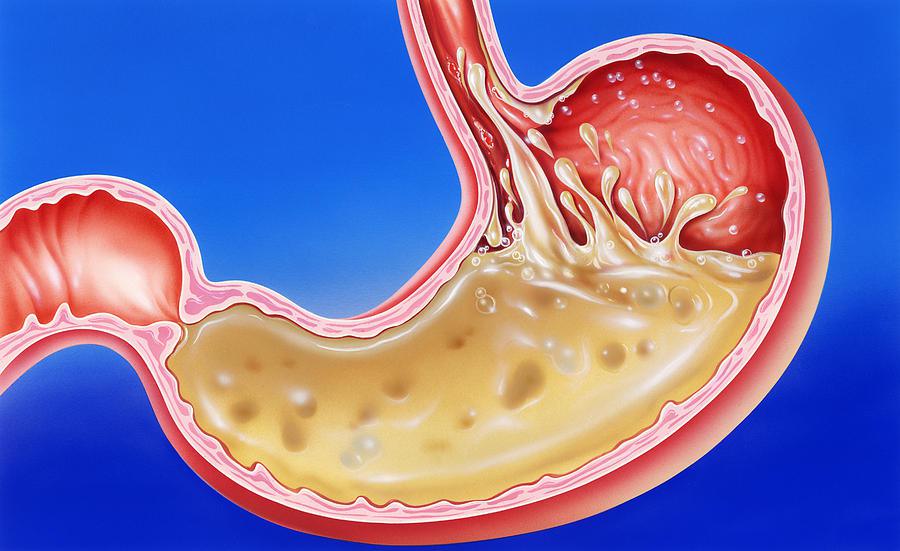
Your Stomach Acid Can Dissolve Metal - Industry Tap

I accidentally swallowed a chicken bone, should I seek medical assistance, or will my stomach acid break it down? - Quora
How long would it take for stomach acid to dissolve a bone lodged within the intestines? - Quora

Question: Can Stomach acid dissolve a bone? (2021)

I accidentally swallowed a chicken bone, should I seek medical assistance, or will my stomach acid break it down? - Quora

Can Stomach Acid Dissolve Metal? Interesting Human Body Facts - YouTube
Shrew-Eating Scientists Show Humans Can Digest Bone | Smart News | Smithsonian Magazine

How Strong Is Stomach Acid? Plus What to Do When Acid Levels Fluctuate
What should I do if I swallow a chicken bone? - Quora
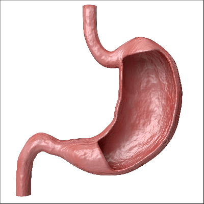
19 Stomach Facts for Kids, Students and Teachers

Hydrochloric Acid vs. Bone total decomposition 🍖 - YouTube

Pin on Fun & Interesting - Did You Know Facts
/GettyImages-585996538-b7755be0a8e849ff93b516e8aa43bfc4.jpg)
Duodenum: Anatomy, Location, and Function
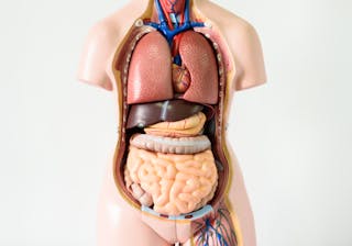
Seven body organs you can live without

Human digestive system - The teeth | Britannica

How Strong Is Stomach Acid? Plus What to Do When Acid Levels Fluctuate

Canine Digestion vs. Human Digestion: What's Different? - Paw Castle

How Strong Is Stomach Acid? Plus What to Do When Acid Levels Fluctuate
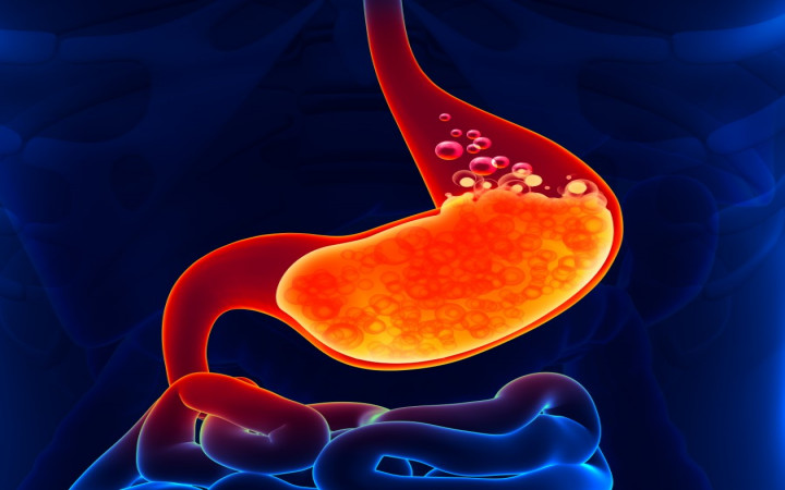
How Powerful Is Stomach Acid? | Wonderopolis

human digestive system | Description, Parts, & Functions | Britannica
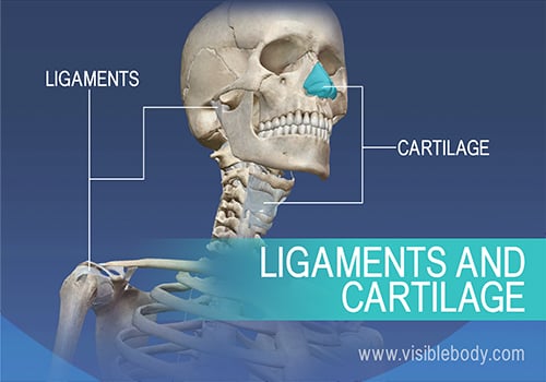
An Anatomy & Physiology Course for Everyone! | Visible Body Learn Site
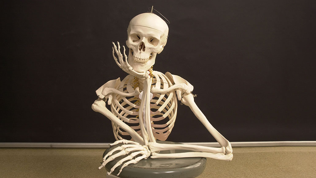
Let's learn about bones | Science News for Students
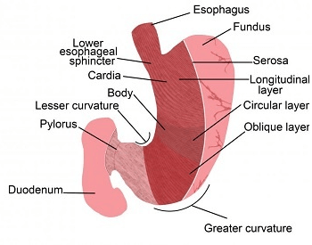
19 Stomach Facts for Kids, Students and Teachers

Fish Bone Stuck in Throat: 9 Ways to Get It Out

human digestive system | Description, Parts, & Functions | Britannica

Can stomach acid dissolve fish bones
Stomach acid can dissolve bones and even metal. What makes corn, chewing gum, and tapeworms unaffected by stomach acid? - Quora
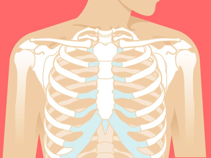
Sternum Anatomy, Location, Function, Pain, Injuries

The digestive and excretory systems review (article) | Khan Academy

Are acidic foods harmful to health?

PDF) Gastric Acid, Calcium Absorption, and Their Impact on Bone Health
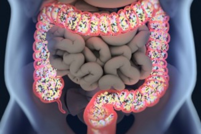
20 Things You Didn't Know About Digestion | Discover Magazine
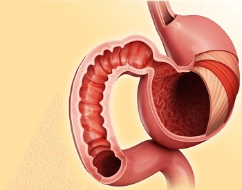
19 Stomach Facts for Kids, Students and Teachers

human digestive system | Description, Parts, & Functions | Britannica

What You Should Immediately Eat When A Fish Bone Gets Stuck In Your Throat - SUCH TV
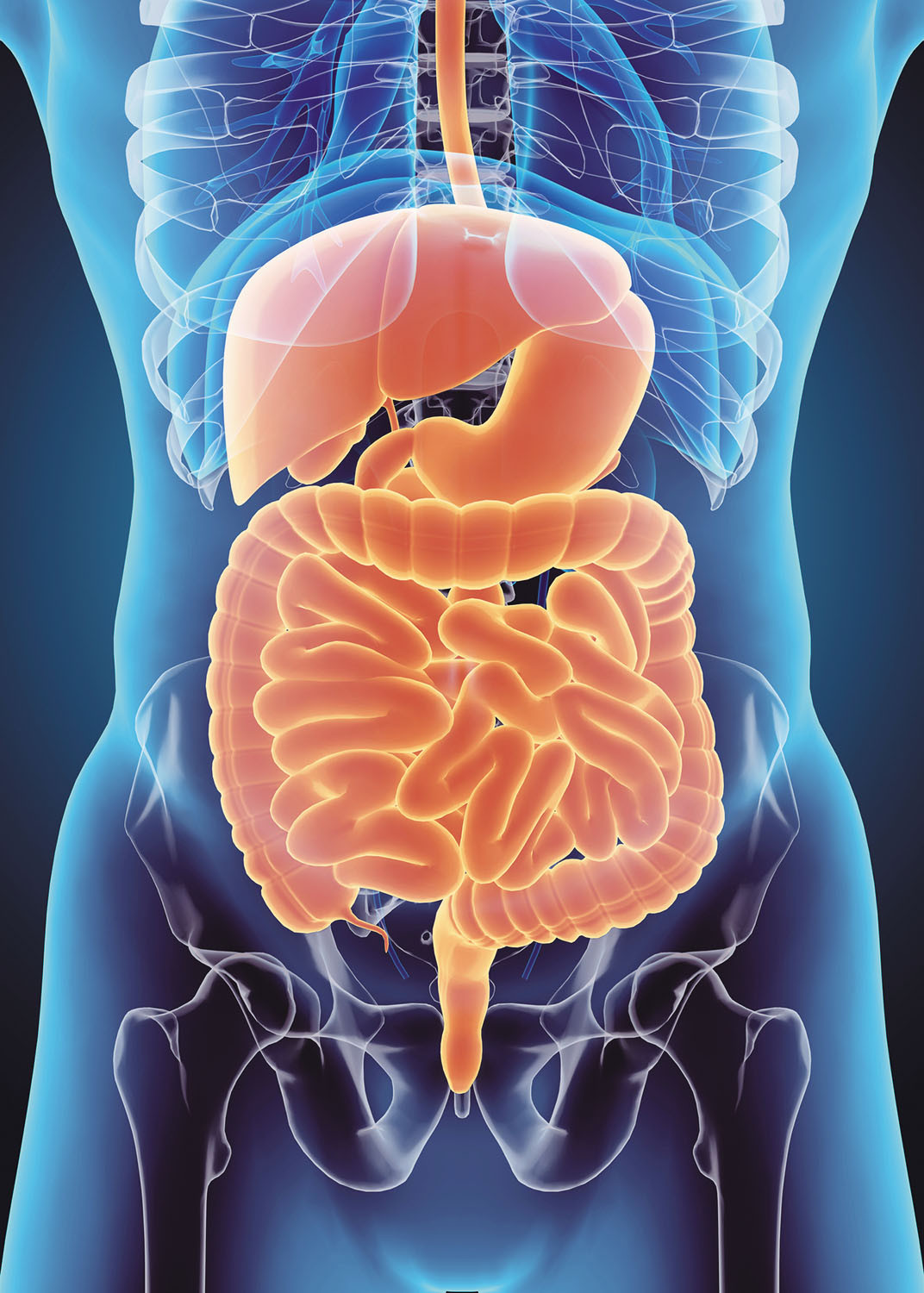
Keep your digestion moving - Harvard Health

Gastrointestinal tract 1: the mouth and oesophagus | Nursing Times
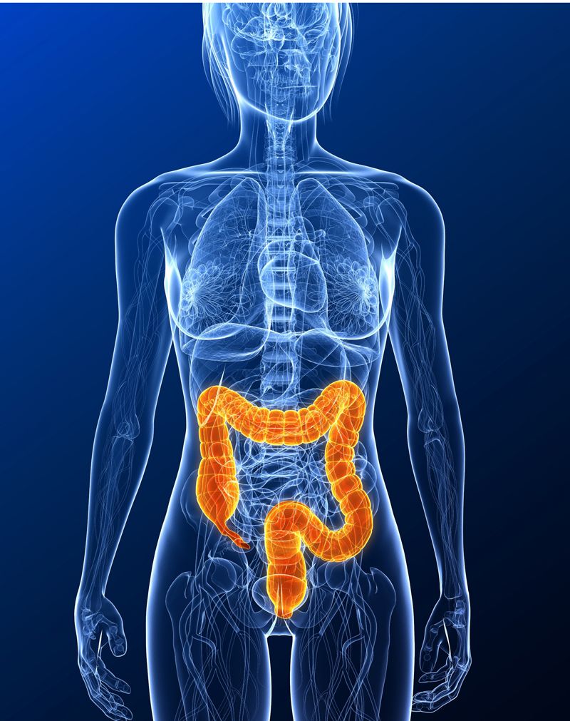
11 Surprising Facts About the Digestive System | Live Science
Posting Komentar untuk "can human stomach acid dissolve bone"